Unprecedented intrauterine procedure saves baby with exposed organs in the United States


A medical team in the United States conducted an unprecedented intervention to repair a serious congenital anomaly in a fetus. The procedure took place in November 2023, when British Maisie Savage was in the 26th week of pregnancy. A resident of London, she had to travel to Texas to provide the specialized care that guaranteed Theo’s survival. The boy came into the world in February 2024 and, at five months old, is showing absolutely healthy growth.
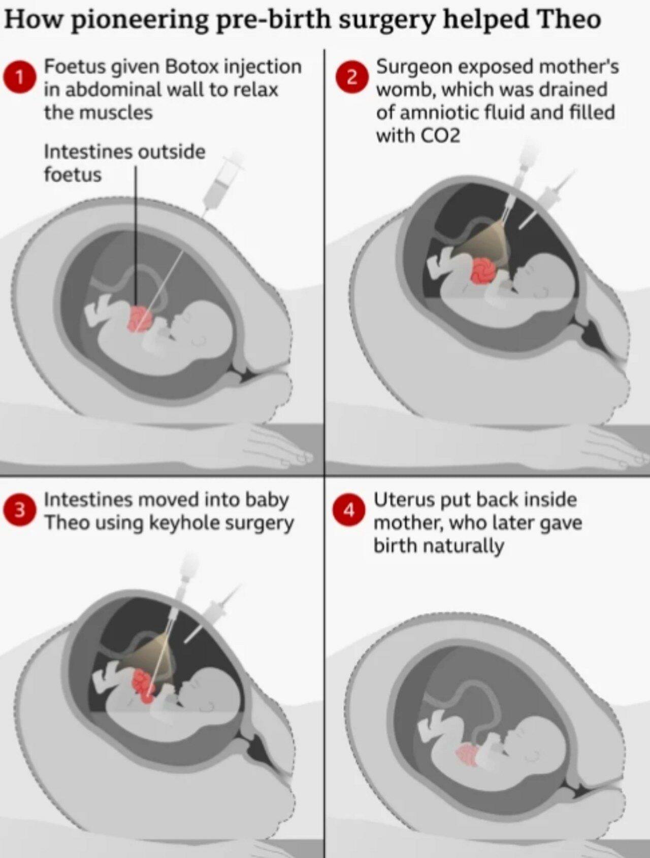
The diagnosis pointed to gastroschisis, a failure in the closure of the abdominal wall that results in the development of the intestine outside the fetus’ body. To correct the problem, specialists partially opened the mother’s uterus and, using video surgery techniques, returned the organs to the child’s abdomen. The operation marked the third global success within this experimental protocol, consolidating Theo as the first patient in the United Kingdom to undergo this highly complex intrauterine correction.
Understand the severity of gastroschisis and the risks for the newborn
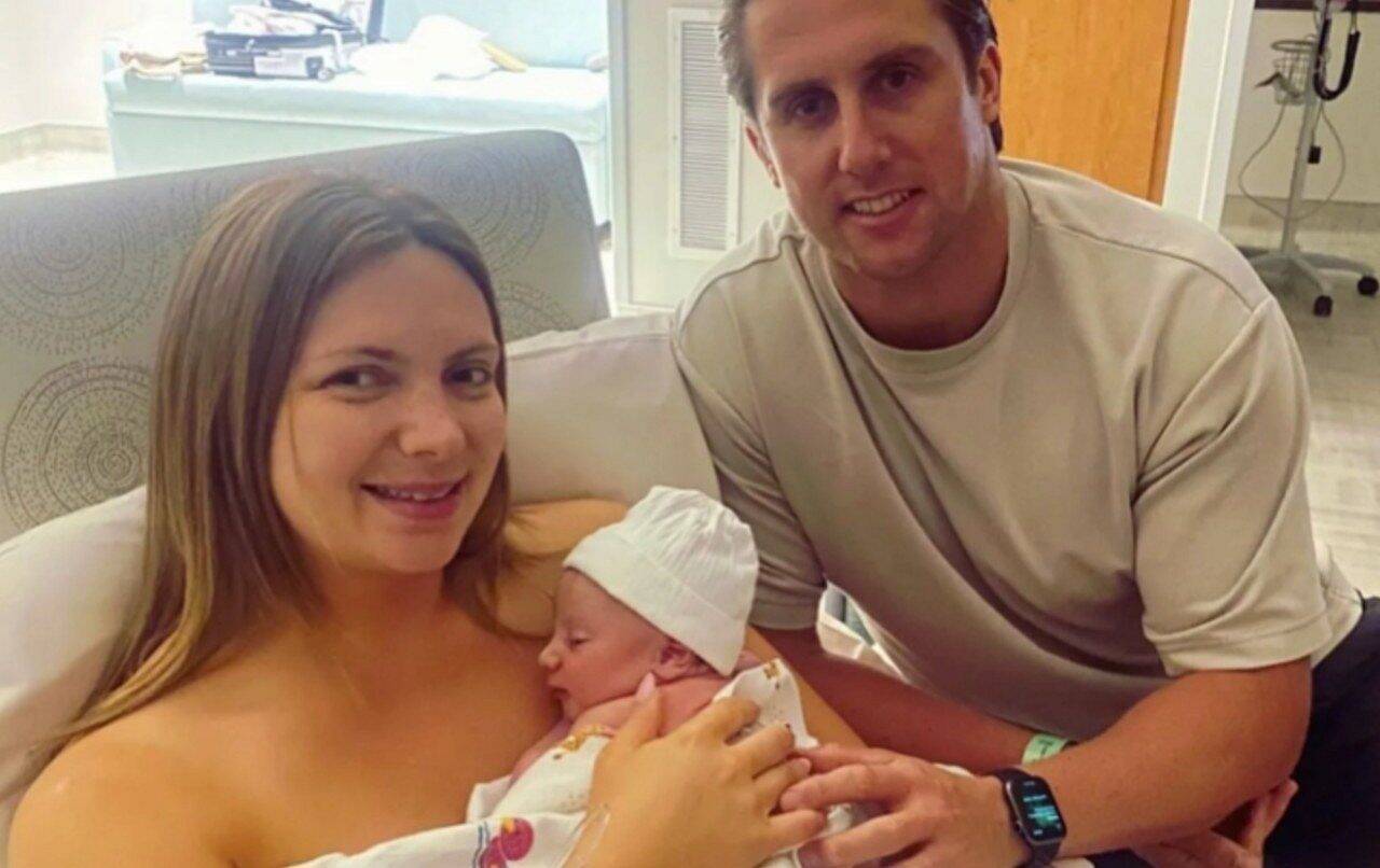
Teachers Maisie, 29, and Josh French, 36, celebrated the positive result of the medical intervention. The mother classifies her son’s survival as an extraordinary feat, being fully aware that the original clinical picture was devastating. In the United Kingdom alone, this anomaly affects one in every three thousand births per year. In milder variations of the disease, newborns often face weeks of hospitalization in intensive care units, depending on intravenous feeding until they undergo traditional corrective surgeries.
However, severe conditions like the one Theo faced bring extreme complications. Prolonged exposure of organs to amniotic fluid causes a severe inflammatory process, damaging tissues even before birth. Because of this, conventional postpartum treatment may require a series of aggressive interventions for the child’s survival:
- Up to half a year of continuous hospitalization in a neonatal intensive care unit.
- Multiple visits to the operating room to try to gradually reposition the intestine.
- Exclusive dependence on tube or intravenous nutrition for up to two years.
- Need for a complete intestinal transplant in the most serious scenarios.
During the first months of pregnancy, the family was unaware of any irregularities in the formation of the fetus. The father recalls that receiving the news of the malformation at the 20-week morphological examination generated paralyzing fear. Four weeks later, upon confirming that it was a complex and much more dangerous variation, doctors at Great Ormond Street Hospital (GOSH), based in the English capital, transferred monitoring to Texas Children’s Hospital, located in the city of Houston.
The international journey in search of a pioneering treatment
At the North American institution, a multidisciplinary group of pediatric and fetal surgeons worked on a clinical study focused precisely on the prenatal correction of this anomaly. The command of the team was in the hands of doctor Michael Belfort, a professional who already had a history of pioneering operations inside the uterus to treat cases of spina bifida. He took direct responsibility for applying the new technique to the British baby.
The medical team informed Maisie of the need for an immediate trip to the United States, where she would have to remain until the moment of birth. Before crossing the Atlantic, protocol required a strategic stop at Leuven Hospital, in Belgium. There, specialists administered an injection of botulinum toxin directly into the abdominal wall of the fetus, passing through the uterus, with the aim of relaxing the muscles and enabling the repositioning of the organs.
Only after this preparatory stage, the couple traveled to Texas, where the definitive surgery took place in November 2023. Josh reports that waiting for the procedure to end was a harrowing experience. The father described those hours of waiting in the hospital as if they were equivalent to an entire week, such was the tension surrounding the risks of the operation.
How surgical intervention worked inside the mother’s womb
The surgical technique required partial exposure of the pregnant woman’s uterus to ensure the correct angle of access. Next, the team drained the amniotic fluid and filled the space with carbon dioxide, creating an expanded area for manipulating the instruments. Using minimally invasive methods, surgeons were able to push Theo’s intestine back into the abdominal cavity, finishing the process with precise sutures on the baby.
The post-operative period left the mother with an abdominal incision twice the size of a traditional cesarean section. The big difference is that, unlike women who undergo a surgical birth, she needed to maintain the pregnancy for another 14 weeks. The normal movements of the fetus caused intense pain in the area of the stitches, making the recovery period quite exhausting, although she guarantees that all the sacrifice was worth it.
The success of the intervention allowed the pregnancy to reach the ideal time, culminating in a normal birth in February 2024, still in Texas. The director of obstetrics and gynecology at the American hospital, Michael Belfort, celebrated the positive outcome. The specialist highlighted the absence of complications both in the fetal phase and after birth, classifying the mother’s performance and the baby’s resistance as determining factors for the extraordinary result.
The Future of Fetal Operations and Baby’s Surprising Recovery
Following the case closely, the head of regenerative medicine at University College London and surgeon at GOSH, Paolo de Coppi, plans to implement this technique in the British healthcare system. The professor awaits the conclusion and definitive success of clinical trials so that the procedure becomes a viable alternative for a much larger volume of pregnant women in the future.
The exchange of knowledge between institutions also generates optimism among the American team. Belfort believes that the union of forces between hospitals in Houston and London creates an unbeatable center of excellence. The doctor points out that the intrauterine environment has consolidated itself as the new frontier of surgical innovation, paving the way for complex cardiac and pulmonary interventions to be carried out even before patients are born.
Already settled back at home in England, the mother reports that the boy has excellent developmental milestones. She highlights that medical intervention managed to nullify what would have been a beginning of life marked by suffering and prolonged hospitalizations. Today, Theo interacts and grows with the same vitality as any other child his age.
The father reinforces his feeling of gratitude for the international support network that saved his son’s life. He highlights the exceptional welcome provided by all professionals involved, from the initial team in London to the group of experts led by Belfort in the United States. Humanized care transformed a period of extreme family trauma into a journey with a happy ending, ensuring the full health of the newborn.