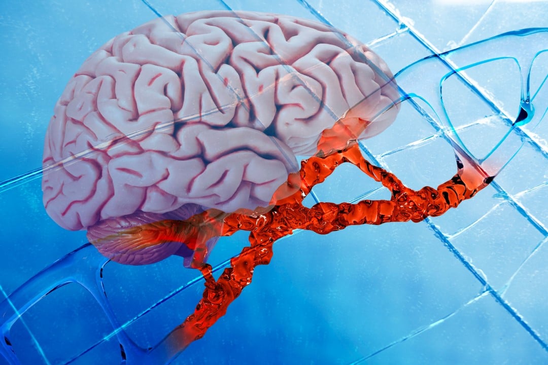
Changes in the genetic material of neurons during the structuring phase of the central nervous system can result in severe functional decline if they are not repaired by the body. A recent investigation conducted by experts from Kyoto University, together with partner research centers, demonstrated that these temporary failures are fundamental to understanding the origin of several neurological pathologies that affect motor coordination and human cognition.
The physical impact of cell migration on the development of the nervous system
The work led by scientist Mineko Kengaku and her group of Japanese academics detailed how the primordial structure of the brain requires extreme physical effort from cells. During the early stages of pregnancy and brain formation, the genetic code of these processing units undergoes literal ruptures due to pressure from the surrounding environment.
Through laboratory tests using animal models, scientists have proven that the neuronal genome is physically crushed as the organ gains volume. This phenomenon occurs because new neurons, newly created from stem cell matrices, need to squeeze through tiny gaps in the biological tissue, generating a mechanical force so intense that it completely deforms the nucleus where the DNA is stored.
In the vast majority of biological scenarios, the body itself can repair these genetic fractures spontaneously and quickly. However, failure in this self-correction mechanism opens the door to chronic dysfunctions and degenerative diseases. The finding was considered so innovative that it gained prominence in the pages of Nature, one of the most respected scientific journals in the world, for offering a missing piece in the puzzle of complex syndromes linked to neurodevelopment.
The extreme journey of neurons to the surface of the cortex
The outer layer of the brain, known as the cortex, is home to billions of connections responsible for conscious thought and voluntary movements. To get there, nerve units are born deep within the organ and travel through an extremely dense cellular network. The medical community already suspected that this trip required colossal mechanical effort, but recording this event live in living organisms was a technical challenge that the Japanese team finally managed to overcome.
By monitoring the exact path of these tiny structures in the brains of growing mice, the researchers mapped each obstacle they faced. They documented that the nuclear capsule suffered severe distortions, culminating in the literal breaking of the DNA double helix when the cell had to cross very rigid anatomical barriers.
In traditional biology, a double-strand break in the genetic code is a major alarm signal, often associated with programmed cell death or the emergence of aggressive tumors. Surprisingly, in the context of brain formation, once the journey was over and the cortex was assembled, the fractures disappeared without a trace, allowing the animals to grow up with perfect cognitive functions.
Crucial differences between the behavior of healthy and tumor tissues
To understand exactly how biology deals with this stress, experts created microscopic artificial mazes, simulating the cramped corridors of the brain. By forcing different cell types to pass through tunnels smaller than their own nuclei, the team noticed completely different reactions:
- In samples of cancerous tissue, mechanical pressure caused the nucleus’s protective membrane to explode, lethally exposing the internal material.
- In samples of healthy nervous tissue, the outer capsule demonstrated impressive flexibility, remaining completely sealed and protecting the internal environment against invaders.
Disruption in diseased cells allowed corrosive substances to invade the nucleus and destroy the genome in a chaotic manner. In neurons, DNA breaking was actually a process controlled by topoisomerase, a protein specialized in untangling genetic strands. This enzyme intentionally cut out parts of the code that didn’t house vital information, relieving physical tension to make it easier to pass through, and patching it all together afterwards.
Long-term consequences of cellular repair failures
The real danger arises when the maintenance system fails. When testing rodents engineered in the laboratory to be unable to fix these genetic breaks, scientists noticed that the puppies appeared healthy in the first few months of their lives. However, over time, the brains of these animals began to suffer from chronic inflammation and loss of essential proteins, resulting in premature aging that directly affected the cerebellum, causing stumbling and severe difficulty in locomotion.
Given this robust evidence, the project leadership at the Asian institution is already outlining the next steps for scientific investigation. The goal now is to decipher the exact impact and evolutionary significance of these lesions programmed into the genome, seeking answers that could, in the future, inspire new treatments to halt the degeneration of the human nervous system.